Description
Features of Sonoscape ProPet 70
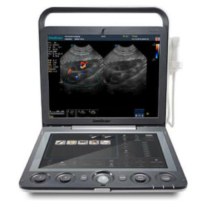
23.8-inch LED Monitor
The ProPet 70 features a 23.8″ full HD LED display for optional, delivering excellent contrast resolution, image clarity and vibrant color in any lighting condition
13.3-inch Tilting Touch Screen
13.3″ anti-glare and anti-fingerprints touch screen with 15 degree rotation
Protective Silicon Overlay of Control Panel
Water-proof and free of animal hairs
Built-in High-capacity Battery
Power management with battery supporting 2 hours continuous scanning per charge in case of power failure
Five Probe Sockets
To save clinicians valuable time and energy by relieving the trouble of changing transducers frequently
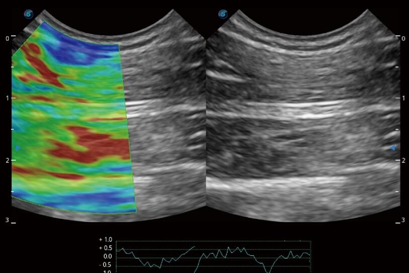
Strain Elastography
Offers a real-time tissue stiffness assessment displayed as a color map to detect potential abnormalities within normal tissues.
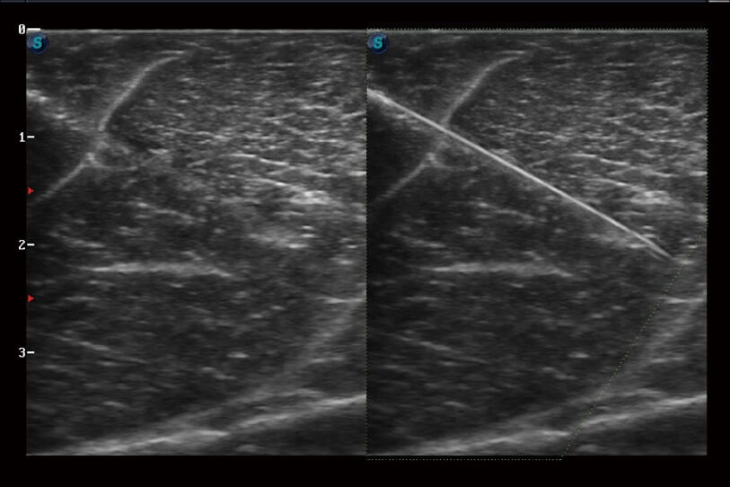
Vis-Needle
Enhanced needle visualization technology reveals needle location within animal anatomy with no distortion when performing interventions like nerve blocks and tissue biopsies.
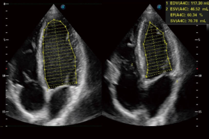
Auto EF
Automatic ejection fraction calculation based on left ventricular wall tracing and Simpson’s rule saves time and efforts compared with manual measurement.
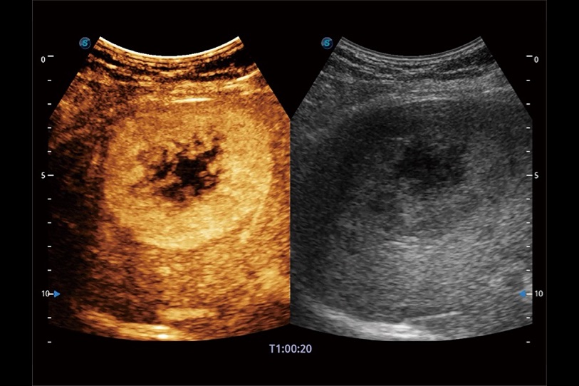
Contrast Enhanced Ultrasound
The non-linear contrast enhanced ultrasound imaging makes full use of harmonic and fundamental signals to give a more enhanced image of difficult-to-view blood flow. Provides a color coded parametric view, indicating the uptake time of contrast agents in different perfusion phases to better differentiate tissues.
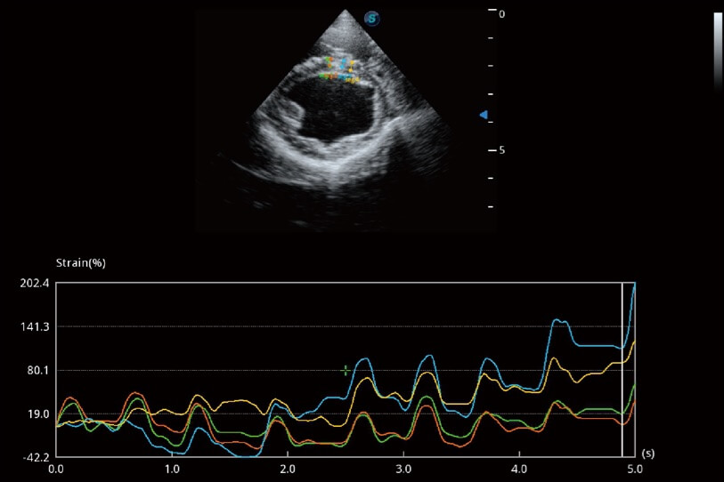
MQA
Precise left ventricular wall motion detection with globally 2D speckle patterns tracking provides accurate quantitative analysis including strain, strain rate, displacement, velocity, etc. on myocardial walls.
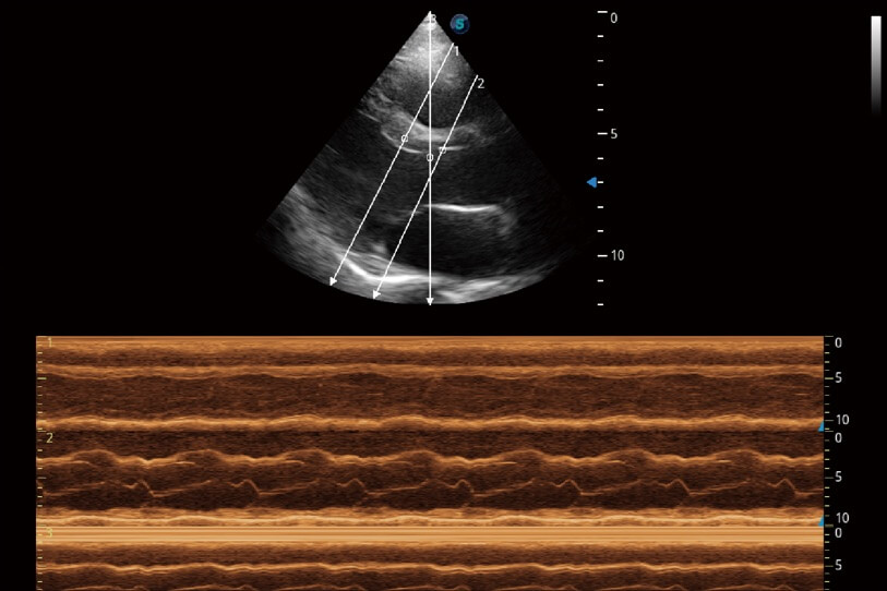
AMM
Collects data with up to three sampling lines at one time to implement detailed assessment on wall motion. It greatly improves the reproducibility and accuracy of left ventricular measurement.